Optical Tweezers
Light exerts force on matter onto which it falls; this was discovered by Maxwell in 1873. There are excellent modern reviews[1],[2] readily available and some details of the contributing forces[3] that trap the biological particles in optical tweezers.
The fact is that light exerts only a miniscule pressure on illuminated objects is part of the difficulty of achieving sufficient containment forces; it is necessary to correctly analyze this phenomenon in some detail to ensure it is strong enough to hold a particle of a molecule in place for examination at leisure. The resulting experimental systems are complicated but their design basis starts with consideration of the energy and momentum of a photon, E = hv, p = E/c = hv/c respectively where c is the speed of light. The important thing is that light carries momentum and anytime it bounces off a surface at some inclination, a corresponding reaction force in the opposite direction accompanies the momentum change. The inset diagram below represents a simple view of what is occurring to a small particle in the beam; scattered photons produce a radial gradient force that tends to push the particle back into the middle of a laser beam source of photons. In addition laser beams have a bell curve (“Gaussian”) profile of optical intensity so they are more intense at their centerline. This too creates net forces that push the particle towards the center of the beam.
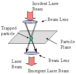
With this arrangement you would expect to accelerate particles in the direction of the centerline of the original beam; however, the optics are arranged in a converging/diverging beam geometry. This puts a restoring force on any trapped particle and confines it to the plane defined by the optics. In addition, the laser beam carries an intense electric field caused by the passage of the photons. This electric field induces dipoles on susceptible molecules and the charge distribution so produced also confines the molecules in the laser beam.
As a result of these combined force fields, optical tweezers have been used to trap dielectric spheres, viruses, bacteria, living cells, organelles (i.e., substructures of cells), small metal particles, and even strands of DNA. Applications include confinement and organization (e.g. for cell sorting), tracking of movement (e.g. of bacteria), application and measurement of small forces, and altering of larger structures (such as cell membranes). Two of the main uses for optical traps have been the study of molecular motors and the physical properties of DNA. In both areas, a biological specimen is biochemically attached to a micron-sized glass or polystyrene bead that is then trapped[2].
Footnotes and References
[1]. http://www.iscpubs.com/articles/al/a0111bro.pdf
[2]. http://www.stanford.edu/group/blocklab/Optical Tweezers Introduction.htm
[3]. http://www.nbi.dk/~tweezer/introduction.htm
Contact: koskyp@union.edu
Self-Assembly from Condensed Phases | Department of Physics & Astronomy | Union College Home Page
Self-Assembly from the Gas Phase by Bottom-Up Technology © 2003 Kosky.
Last Updated: 08/05/05 01:12 PM